Event

Listen to world-leading experts in imaging

About

The EPFL Seminar Series in Imaging

Imaging plays a central and ever-increasing role in science and engineering. From the nano to the macro scale, it allows us to capture, quantify, and visualize physical phenomena with unprecedented resolution in both space and time.

It is also the interdisciplinary discipline par excellence. From sample preparation to optical design and image processing, imaging workflows nowadays require the convergence of numerous skills and expertise.

Mindful of this “imaging sweet-spot”, the EPFL Center for Imaging aims at bringing together the best from worldwide experts in imaging through a series of high-visibility talks with interdisciplinary appeal.

September 23, 2024
September 29, 2024

The Scientific Images Exhibition 2024

Do you have a passion for unraveling and witnessing the breathtaking beauty of scientific elements and phenomena? If so, we invite you to share your scientific vision through the lens of your camera, microscope, telescope, computer, etc.!
Submission deadline:
What's next

Upcoming seminars

February
27
,
2025
,
17:00

Revolutionizing Cellular Imaging: Harnessing Label-Free Flow Cyto-Tomography for Advanced Suspended Cell Analysis

Prof. Pietro Ferraro, IMM CNR, Italy
SV 1717
Registration required
Abstract:

Abstract:

This lecture will explore the innovative application of label-free flow cyto-tomography in the study of suspended cells. Traditional methods of cellular analysis often rely on labeling techniques that can alter or obscure native structures, limiting the accuracy of observations. Flow cyto-tomography, however, provides a powerful, non-invasive alternative for visualizing and quantifying the internal architecture of cells in suspension. By combining the principles of flow cytometry with high-resolution tomographic imaging, this technique offers unprecedented insights into cellular morphology, organelle organization, and quantification of subcellular structures. The lecture will cover the underlying technology, its applications in biomedical research, and its potential to advance our understanding of cellular function in health and disease. Looking ahead, this approach could pave the way for novel diagnostic tools and therapeutic strategies, opening new frontiers in personalized medicine and cellular engineering.

Bio:

Director of Research at the National Research Council (CNR) of Italy, affiliated with the Institute of Applied Sciences and Intelligent Systems "Eduardo Caianiello" (ISASI). His core expertise lies in the fields of holography and microscopy, with a particular focus on developing and applying cutting-edge imaging techniques. He is renowned for his contributions to digital holography, holographic microscopy, and related optical methods. His research frequently explores applications in diverse areas, including biomedicine, materials science, and cultural heritage. He often works on advanced optical systems for visualizing and analyzing microscopic structures, pushing the boundaries of what's possible in high-resolution and three-dimensional imaging. In recent years his research has focused on intelligent systems and he has in fact developed the so-called Lab on a Chip, Founder of the Institute of Applied Sciences and Intelligent Systems of the CNR in 2016 and author of over 600 scientific publications, in 2020 he was awarded the international "SPIE-Dennis Gabor Award".

May
08
,
2025
,
17:00

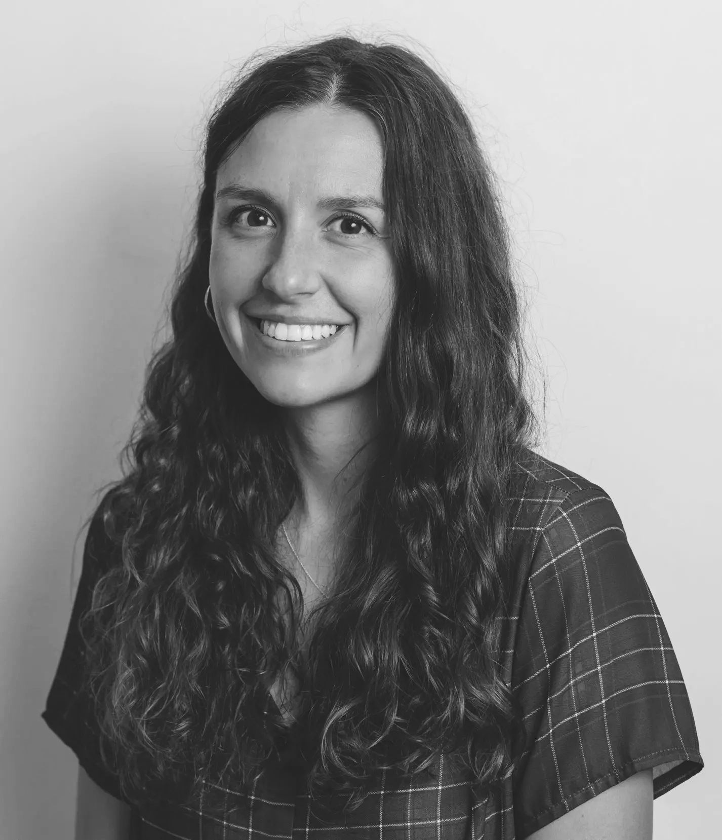
Quantifying Quality in Fluorescence Microscopy

Prof. Siân Culley
SV 1717
Registration required
Abstract:

Fluorescence microscopy is a central technique for quantifying the spatiotemporal distribution of cellular compartments. However, the ability to accurately retrieve biological information from images depends strongly on the quality of the data. The perceived quality of fluorescence microscopy images can be improved by imaging with higher illumination doses, however this comes at the cost of potentially damaging and altering the behaviour of living samples. To reduce the impact of noise when acquiring at lower illumination densities, image processing techniques have been developed, many of which are based on AI. Here, we interrogate quality metrics which are commonly used to assess the performance of image processing methods. These methods are commonly used in microscopy, but have their origins in computer vision. We show that these metrics report not just image quality, but also other characteristic features of fluorescence microscopy images, which can confound their interpretation. We also quantify how predictive these metrics are of successful downstream image analysis for a variety of common tasks, and discuss how and when these metrics should be best used.

Bio: Siân Culley is a Royal Society University Research Fellow who started her group at King’s College London in 2021. Her interests are in how microscopy methods can be tailored to best fit biological questions, with a particular focus on assessing data quality and reliability. Prior to starting her group she did postdoctoral work with Ricardo Henriques at UCL, developing open source methods for super-resolution fluorescence microscopy.

May
22
,
2025
,
17:00

Developing imaging technologies to search for, discover, and understand ocean life

Dr. Kakani Katija, MBARI USA
BM 5202
Registration required
Abstract:

As lead of the Bioinspiration Lab, Kakani and her group investigates ways that imaging can enable observations of life in the deep sea. By developing novel imaging and illumination tools (e.g., DeepPIV and EyeRIS), automating the classification of underwater visual data using artificial intelligence (FathomNet), and integrating algorithms on vehicles (ML-Tracking) for robotic vehicle missions (e.g., Mesobot, LRAUV) to consistently and persistently observe ocean life, their efforts will increase access to biology and related phenomena in the deep sea. If successful, the Bioinspiration Lab hopes to spark collaborative research and engineering innovations inspired by poorly understood inhabitants living in the least explored habitat on our planet.

Bio: Kakani Katija completed her bachelor's degree in Aeronautics and Astronautics at the University of Washington in 2004. She furthered her studies, earning a Master's in Aeronautics in 2005 at the California Institute of Technology (Caltech) and her Doctorate at Caltech in 2010 in Bioengineering. She served as a Postdoctoral Fellow at the Monterey Bay Aquarium Research Institute.Katija was awarded research fellowships from both the American Society for Engineering Education and the National Science Foundation to conduct graduate research. As a certified research diver, she has conducted field studies in various locations throughout the world, including research completed in 2009 off the coast of the Palau archipelago. The goal of this study was to understand the physics involved in the movement of jellyfish. The science team discovered the jellyfish not only push water into their bells but drag an almost constant flume of water behind them. This discovery led Katija to study how marine life contributes to mixing the ocean. Katija's work also includes understanding how much sea creatures mix fluid in the ocean at rates comparable to winds and tides. Katija now leads the Bioinspiration lab at the Monterey Bay Aquarium Research Institute in Moss Landing, California, where she has developed DeepPIV, a research tool intended to make conducting experiments in ocean habitats less invasive and improve marine research techniques. She has participated in two expeditions on board R/V Falkor - Designing the Future and Designing the Future 2 testing the newly developed technologies on board. Imagery from the use of the DeepPIV used on board Schmidt Ocean Institute's R/V Falkor, is available on SketchFab.

June
18
,
2025
,
17:00

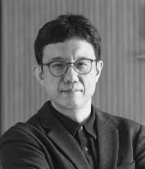
Meta Devices for Photonics and Quantum

Din Ping Tsai, University of Hong Kong
SV 1717
Registration required
Abstract:

Specially designed meta-structure components can mass-produce artificial nano-array structures through semiconductor microelectronics fabrication procedures, and can manipulate the phase, polarization, and amplitude of electromagnetic waves. Meta-devices can meet a variety of current urgent needs, such as novel functions, lightweight, small size, higher efficiency, better performance, broadband operation, lower energy consumption, compatibility with semiconductor mass processing technology, etc. This talk reports on the design, manufacturing and novel applications of optical meta-devices, such as achromatic meta-lens, advanced imaging, intelligent sensing, nonlinear generation of vacuum ultraviolet light, medical and biological imaging, 6G communications, tunable meta-structures, high-dimensional quantum light source, etc. We believe that meta-devices have opened up a new avenue for future developments of next-generation devices in fields such as micro-robotic vision, autonomous driving, vehicle sensors, virtual and augmented reality, personal miniature security systems, bio-medical devices, advanced medical care, and quantum information technology, etc.

Bio: Din Ping TSAI He is a Chair Professor of Dept. of Electrical Engineering, City Univ. of Hong Kong. He is Fellow of AAAS, APS, COS, EMA, IEEE, JSAP, NAI, OSA, SPIE, and AAIA, respectively. He is Member of the International Academy of Engineering (IAE), and Academician of the Asia-Pacific Academy of Materials (APAM) and Hong Kong Academy of Engineering (HKAE), respectively. He is the author of 388 refereed papers. He was granted 69 patents for 45 innovations. He was invited speaker for international conferences more than 355 times (31 Plenary Talks, 66 Keynote Talks). He received more than 40 prestigious recognitions and awards, including “Mozi Award” from the International Society of Optical Engineering (SPIE) in 2018; “Global Highly Cited Researchers,” by Web of Science Group (Clarivate Analytics) in 2020 and 2019, respectively; China’s Top 10 Optical Breakthroughs in 2020 and 2018, respectively; and 2024 Frontiers of Science Award; etc.

September 23, 2024
September 29, 2024

The Scientific Images Exhibition 2024

Do you have a passion for unraveling and witnessing the breathtaking beauty of scientific elements and phenomena? If so, we invite you to share your scientific vision through the lens of your camera, microscope, telescope, computer, etc.!
Submission deadline:
Past seminars

Explore our previous events

Review our former seminars and watch recordings if available.

Correlative cryoSEM-CryoNanoSIMS mapping of vitrified biological tissue

By. Dr. Priya Ramakrishna
EPFL

Visualizing mechanical properties in biology using Brillouin microscopy

Dr. Robert Prevedel
European Molecular Biology Laboratory

Deep Learning-enabled Computational Microscopy and Diffractive Imaging

By Prof. Aydogan Ozcan
UCLA

Simultaneous 3D imaging in Biology with Multifocus Microscopy

Prof. Sara Abrahamsson
University of California Santa Cruz (US)

Normalizing Flows and the Power of Patches in Inverse Problems

Prof. Gabriele Steidl
TU Berlin

Future of Bioimaging: Next Generation Instruments & Artificial Intelligence

Prof. Loïc Royer
Institut Curie

Posterior-Variance-Based Error Quantification for Inverse Problems in Imaging

Prof. Thomas Pock
Graz University of Technology

Scanning Ion Conductance Microscopy

By Prof. A. Radenovic & G.Fantner
EPFL

Generative AI, Stable Diffusion, and the Revolution in Visual Synthesis

By Prof. Bjorn Ommer
LMU of Munich

From differential equations to deep learning for inverse imaging problems

By Prof. Carola Bibiane Schönlieb
University of Cambridge

Imaging using X-ray scattering Contrast to Bridge the Nano and Macroscale

By Prof. Marianne Liebi
EPFL and Paul Scherrer Institute

Joint Optimization of Learning-Based Image Reconstruction and K-Space Trajectories for MRI

By Prof. Jeff Fessler
University of Michigan

Integrating Physical and Learned Models

By Prof. Ulugbek Kamilov
Washington University in St. Louis

Modeling Deep Networks: Network Learning for Image Processing

By Prof. Stéphane Mallat
Collège de France

Mapping 3D Nanostructures with X-Ray Ptychography

By Dr. Manuel Guizar-Sicairos
Paul Scherrer Institut, Switzerland Copyright

Beyond the First Portrait of a Black Hole

By Prof. Katie Bouman
California Institute of Technology, USA

3D Imaging of Cells by FIBSEM with Correlation to cryoFLM

By Prof. Harald Hess
HHMI’s Janelia Research Campus, USA

Imaging the Planet for a Sustainable Future

By Prof. Gilberto Camara
Group on Earth Observations (GEO)

End-to-end Learning for Computational Microscopy

By Dr. Laura WALLER
Computational Imaging Lab, UC Berkeley

Imaging: Intelligence on the Nanoscale

By Prof. Gabriel Aeppli
Paul Scherrer Institute, Switzerland

Reconstruction of Cryo-EM Images of Proteins at Atomic Resolution

By Dr. Sjors Scheres
MRC Laboratory of Molecular Biology, Cambridge

Imaging the Unseen: Taking the First Picture of a Black Hole

By Katie Bouman
Caltech, USA

Machine Learning for Bioimage Informatics (AMLD 2020)

By Virginie Uhlmann
EMBL-EBI, UK

Shedding Light on Tumour Evolution

By Prof. Sarah Bohndiek
University of Cambridge, UK

Functional Ultrasound Imaging gcmbvc

By Prof. Mickael Tanter
INSERM and ESPCI Paris, France

Structural Analysis with Cryo-Transmission Electron Microscopy

By Prof. Henning Stahlberg
University of Basel, Switzerland

Imaging: From Compressed Sensing to Deep Learning

By Prof. Yonina Eldar
Weizmann Institute of Science, Israël

Video Understanding and Robotics Manipulation (AMLD 2020)

By Cordelia Schmid
INRIA, France

Ultrastructural Expansion Microscopy

By Prof. Paul Guichard
University of Geneva, Switzerland

On Micro and Nano Imaging ghcmcm

By Prof. Jacques Dubochet
Nobel Laureate in Chemistry, 2017

Reflection Matrix Approaches for Imaging

By Mathias Fink
ESPCI Paris, France

Super-Resolution in Diffraction Microscopy

By Anne Sentenac
Institut Fresnel, France

On Instabilities, Paradoxes and Barriers in Deep Learning

By Prof. Anders Hansen
University of Cambridge, UK

Content Aware Image Restoration for Light and Electron Microscopy

By Florian Jug
Max Planck Institute, Dresden, Germany

Exploring and Explaining: Leveraging data visualization for research, communication and public health

By Prof. Helena Jambor
University of Applied Sciences of the Grisons

Diffusion Models for Computational Imaging Problems
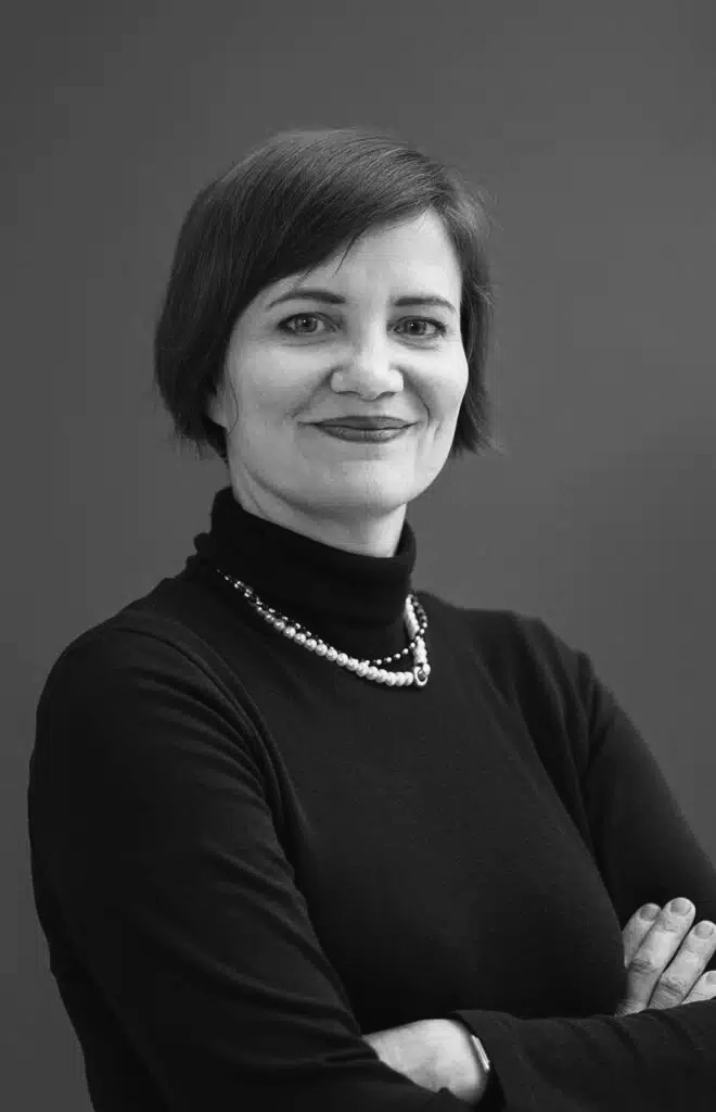
Prof. Jong Chul Yue from KAIST, Korea
SV 1717
Contact

A question on the Center?
Get in touch!

Contact us if you have any questions or require support in the field of imaging.